CryoLetters Volume 46 - Issue 1
CryoLetters 46 (1), 1–13 (2025)
© CryoLetters, editor@cryoletters.org
doi.org/10.54680/fr25110110112
PERSPECTIVE: Production and cryopreservation of 3D cultures
Nataliia Moisieieva, Olga Gorina*, Anton Moisieiev and Olga Prokopiuk
- Institute for Problems of Cryobiology and Cryomedicine of National Academy of Sciences of Ukraine, 23, Pereyaslavska Str., Kharkiv 61016, Ukraine
*Corresponding author’s E-mail: ogorina2603@gmail.com
Abstract
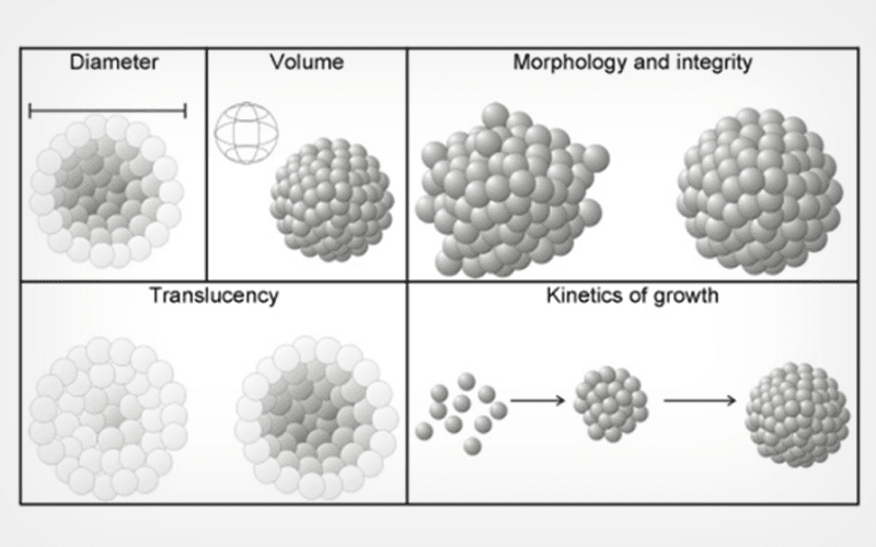
Three-dimensional (3D) culture systems, which include spheroids (SPs), provide a unique platform for studying complex biological processes in vivo and for enhancing the capabilities of in vitro test systems. Their uniqueness lies in the 3D organization of cells and in the reproduction of complex intercellular interactions, similar to those in native tissues and organs. These "mini-organs" can be used for fundamental research, tissue-engineering constructs, development of preclinical models for testing pharmacological drugs, etc. Important and current issues regarding SPs involve improving methods for their production and cryopreservation. Solving these issues will expand the range and effectiveness of their use in tissue engineering. Here, we describe the authors' research and experience on factors influencing the formation of SPs, which can enhance the understanding of their correct application and standardization. A crucial aspect of this review is the information on applying theoretical approaches based on physico-mathematical calculations to improve the quality of existing cryopreservation protocols for SPs.
Keywords: cryopreservation; spheroid; parameters affecting the spheroid formation; theoretical models
CryoLetters 46 (1), 14-21 (2025)
© CryoLetters, editor@cryoletters.org
doi.org/10.54680/fr25110110312
Cryopreservation and assessment of Honamli and Hair buck semen using resveratrol
Muhammed Enes İNANÇ*1, Şükrü GÜNGÖR1, Feyza Nur MART2, Mine HERDOĞAN2, Hasan Ali ÇAY1,2, Durmuş KAHRAMAN2, Fırat KORKMAZ1, Barış Atalay USLU3 and Ayhan ATA1
- Department of Reproduction and Artificial Insemination, Faculty of Veterinary Medicine, Burdur Mehmet Akif Ersoy University, TR-15200, Burdur-Türkiye
- Department of Reproduction and Artificial Insemination, Burdur Mehmet Akif Ersoy University Health Science Institue, TR-15200, Burdur-Türkiye
- Department of Reproduction and Artificial Insemination, Faculty of Veterinary Medicine, Cumhuriyet University, TR-58140, Sivas-Türkiye
*Corresponding author’s E-mail: enesinanc@hotmail.com (ORCID: 0000-0001-6954-6309)
Abstract
Background
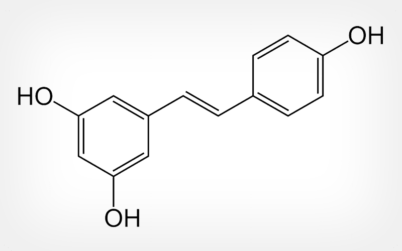
Resveratrol (Res) (3,5,4′-trihydroxystilbene) is a natural polyphenol that exhibits important biological activities.
Objective
To assess the effects of resveratrol (Res) on freeze-thawed survival of semen from Honamli and Hair Bucks.
Materials and methods
Six bucks, aged 2–3 years (three from each breed), were included in the study. Semen was collected from each breed and mixed separately after removing seminal plasma. The mixed semen was diluted with different Res concentrations (0 μM as control, 25 μM, 50 μM, 100 μM, 500 μM, and 1 mM) in Tris diluent and subjected to cryopreservation in liquid nitrogen vapor and frozen. After thawing, the samples were evaluated for motility and some spermatologic quality parameters by flow cytometry.
Results
Data were analyzed separately for Honamli and Hair breeds. The results showed that the Res 1 mM group had the lowest motility in all assessments (P<0.05). However, no significant differences were observed between the other Res and control groups (P>0.05). In terms of apoptosis, Hair bucks exhibited a statistically significant difference in late apoptotic parameters, with the control showing the highest values (P<0.05). The Res 25 μM group (similar to the control group) showed lower mitochondrial oxidative stress than the Res 1 mM group (P<0.05).
Conclusion
Res at a dose of 1 mM did not protect most sperm functional and biochemical parameters except for apoptosis and performed worse than the control group. When all parameters are evaluated collectively, concentrations lower than 1 mM should be used for freezing Honamli and Hair Buck semen with resveratrol.
Keywords: buck; cryopreservation; resveratrol; semen; spermatological parameters
CryoLetters 46 (1), 22-30 (2025)
© CryoLetters, editor@cryoletters.org
doi.org/10.54680/fr25110110512
Effect of in vitro culture as a sperm selection method before single sperm cryopreservation of testicular sperm from individuals with azoospermia
Behnam Maleki1,2, Serajoddin Vahidi1,3, Lida Gholizadeh1,2, Keivan Lorian1
and Azam Agha-Rahimi1*
- Research and Clinical Center for Infertility, Yazd Reproductive Sciences Institute, Shahid Sadoughi University of Medical Sciences, Yazd, Iran
- Infertility Center, Mazandaran University of Medical Sciences, Sari, Iran
- Andrology research center, Yazd Reproductive Sciences Institute, Shahid Sadoughi University of Medical Sciences, Yazd, Iran
*Corresponding author’s E-mail: 63rahimi@gmail.com
Abstract
Background
Single sperm cryopreservation (SSC) is a method that preserves the limited number of spermatozoa in testicular sperm. But testicular spermatozoa are characterized with low movement, which is not ideal for sperm selection before SSC.
Objective
This study was designed to investigate in vitro incubation (IVC) as a sperm selection technique before SSC on biological factors of testicular spermatozoa.
Materials and methods
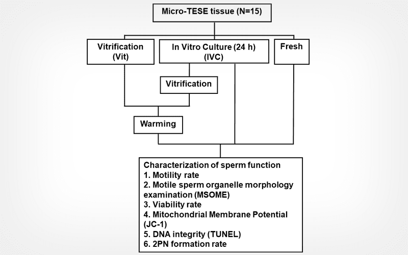
Testicular tissue was obtained from 15 azoospermia men. One part of the testicular samples was used as a Control group, which was assessed fresh. One portion was cryopreserved by a vitrification (Vit) method and the two other portions were in vitro cultured for 24 h, with (IVC-Vit) or without (IVC) vitrification. Sperm motility, viability, morphology, DNA fragmentation and mitochondrial membrane potential were evaluated.
Results
Sperm motility and viability were better maintained in the IVC-Vit group compared to the Vit group (P=0.04 and P= 0.003, respectively). Sperm morphology, the fresh, Vit, IVC, and IVC-Vit groups all showed similar results (P >0.05). Mitochondrial activity was significantly lower in the Vit group compared to the Control fresh group (P = 0.0001). The IVC group had a significantly higher DFI as compared to the Control (P <0.0001). Compared to the IVC group, the IVC-Vit sperm had a significant increase in DFI (P= 0.0009). There was a statistically significant difference between post warm DFI of the Vit group and IVC-Vit group (P <0.0001).
Conclusion
IVC as a sperm selection method increased motility and viability of testicular spermatozoa before single sperm vitrification. As DNA fragmentation increased by this technique, this method is not ideal for selecting viable sperm.
Keywords: azoospermia; cryopreservation; DNA fragmentation; in vitro culturing.
CryoLetters 46 (1), 31-40 (2025)
© CryoLetters, editor@cryoletters.org
doi.org/10.54680/fr25110110712
Effect of carnitine on Hariana bull spermatozoa function after cryopreservation
Brijesh Kumar Yadav1*, Jitendra Kumar Agrawal1, Kavisha Gangwar2, Dilip Kumar Swain3, Brijesh Yadav3, Anuj Kumar1, Vikas Sachan1, Mukul Anand3, Brijesh Kumar4 and Atul Saxena1
- Department of Veterinary Gynaecology & Obstetrics,
- Department of Veterinary Pathology,
- Department of Veterinary Physiology, College of Veterinary Science & Animal Husbandry, Deendayal Upadhayaya Pashu Chikitsa Vigyan Viswavidyalaya Evam Go Anusandhan Sansthan, Mathura, 281001, Uttar Pradesh, India
- ICAR-IVRI Izatnagar, Bareilly, 243122, Uttar Pradesh, India
*Corresponding author’s E-mail: brijeshyadav1090@gmail.com
Abstract
Background
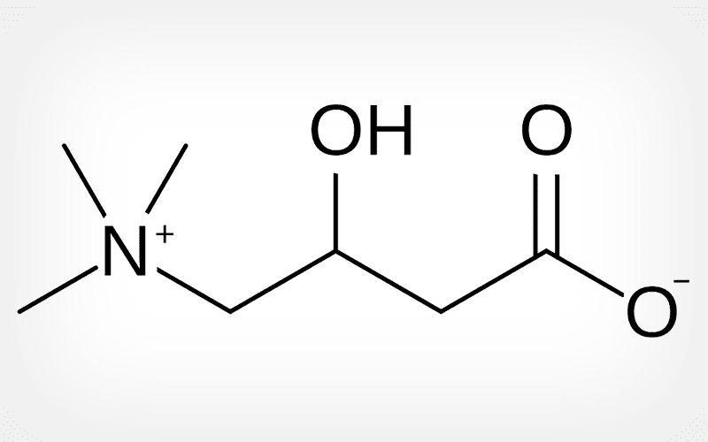
Carnitine reduces reactive oxygen species-induced apoptosis and DNA fragmentation through its antioxidant effect.
Objective
To investigate the effect of carnitine on capacitation, mitochondrial activity, acrosomal integrity, reactive oxygen species (ROS), membrane fluidity, and DNA fragmentation during the cryopreservation of Hariana bull spermatozoa.
Materials and methods
Thirty-two semen ejaculates were obtained using artificial vagina (AV) from four seemingly healthy Hariana bulls. Following dilution, the diluted semen samples were split into four aliquots: Group I, the control, included no carnitine; Groups II, III, and IV were the aliquots that contained carnitine supplements of 2.5, 5, and 10 mM, respectively. These four diluted semen samples were then processed immediately for freezing and equilibration.
Results
Regarding post-thaw sperm parameters, such as viability, motility, velocity parameters, capacitation status, mitochondrial activity, acrosomal integrity, reactive oxygen species (ROS), membrane fluidity, and DNA fragmentation, Groups II and III, containing 2.5 mM and 5 mM carnitine respectively, had significantly (P<0.05) improved parameters compared to the Group I (control). At 5 mM, there was a substantial (P<0.05) decrease in early apoptotic-like alterations in sperm cells, accompanied by a greater population of sperm cells with high mitochondrial membrane potential.
Conclusion
Carnitine has been shown to have cryoprotective properties in semen extenders. For improved post-thaw sperm quality, carnitine may be added to a Hariana bull semen extender at a dose of 5 mM.
Keywords: antioxidant; carnitine; cryopreservation; DNA fragmentation; flow-cytometry
CryoLetters 46 (1), 41-46 (2025)
© CryoLetters, editor@cryoletters.org
doi.org/10.54680/fr25110110212
Effects of cryolipolysis on subcutaneous adipose tissue of adult women: immunohistochemical analysis
Chistiane Rodrigues Tofoli Palauro1, Priscila da Silveira Palmeira Silva Daumas2, Eneida de Morais Carreiro3, Felipe Pimentel de Almeida4, Igor Lustosa Dias5 and Patrícia Froes Meyer3*
- Centro Universitário de Vila Velha, Vila Velha, Brazil
- Universidade Estácio de Sá - UNESA, Brazil
- International Research Group - IRG, Natal, Brazil
- Estácio de Sá University - Fortaleza, Brazil
- Universidade CEUMA, Maranhão, Brazil
*Corresponding author’s E-mail: patricia.froesmeyer@gmail.com
Abstract
Background
The skin, the largest organ in the human body, is composed of complex layers that include subcutaneous adipose tissue. Understanding the characteristics of this skin structure is essential to optimize therapeutic interventions, such as cryolipolysis, aiming for more effective and personalized results.
Objective
To evaluate the immunohistochemical effects of skin tissue in adult women undergoing cryolipolysis.
Materials and methods
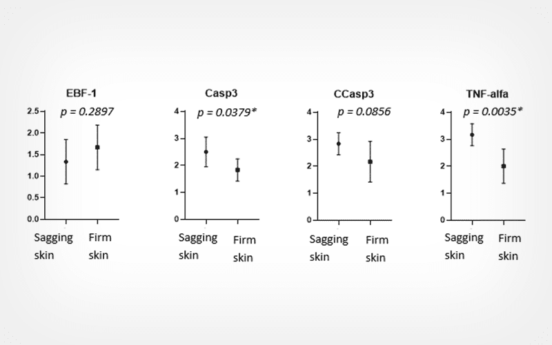
We carried out an experimental and blind study with immunohistochemical analysis in women with localized abdominal fat, categorized based on the constitution of the skin as flaccid or firm according to the Investigator Assessment Skin Laxity Scoring System scale. Participants were randomized before undergoing the cryolipolysis procedure. Forty-five days after the procedure, they underwent abdominoplasty, with collection of biological material. We evaluated the inflammatory markers EBF-1, TNF-alpha, and CD68, as well as Caspase 3, cleaved Caspase 3, apoptotic BCL2, Ki-67 for fibroblast proliferation, and FIS1 for mitochondrial proliferation.
Results
Six women were included, divided into two groups; three women with loose and three with firm skin. We observed that after cryolipolysis, the group with flaccid skin showed higher expression of the Casp3, TNF-alpha, BCL2, and FIS1 markers compared to those with firm skin.
Conclusion
Cryolipolysis may act differently according to tissue morphology, suggesting that its apoptotic response is more pronounced in the group with flaccid skin.
Keywords: abdominoplasty; adipocytes; cryotherapies; esthetics; weight loss
CryoLetters 46 (1), 47-56 (2025)
© CryoLetters, editor@cryoletters.org
doi.org/10.54680/fr25110110412
Non-permeable cryoprotectants’ influence on fibroblast slow freezing in six-banded armadillo
Denilsa Pires Fernandes1, Érika Almeida Praxedes1, João Vitor da Silva Viana1, Maria Valéria de Oliveira Santos1, Carlos Iberê Alves Freitas2 and Alexsandra Fernandes Pereira1*
- Laboratory of Animal Biotechnology, Federal Rural University of Semi-Arid (UFERSA), Mossoro, RN, Brazil
- Laboratory of Studies in Immunology and Wild Animals, UFERSA, Mossoro, RN, Brazil
*Corresponding authors’ E-mail: alexsandra.pereira@ufersa.edu.br
Abstract
Background
There is a crucial need to develop appropriate cryopreservation solutions so that somatic resource biobanks of wildlife can be established.
Objective
Here, we propose a cryopreservation protocol to optimize the preservation of skin-derived fibroblasts from six-banded armadillos (Euphractus sexcinctus Linnaeus, 1758) by comparing different concentrations of fetal bovine serum (FBS) in the absence or presence of sucrose as non-permeable cryoprotectants.
Materials and methods
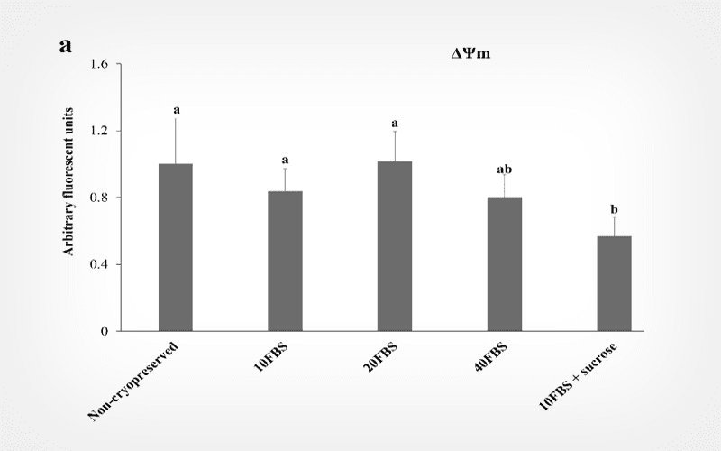
Cells were cryopreserved by slow freezing with different solutions containing Dulbecco’s modified Eagle’s medium (DMEM) with 10% dimethyl sulfoxide (DMSO), varying concentrations of FBS (10, 20 and 40%) without or with 0.2 M sucrose, totaling six comparison groups. Cells not subjected to cryopreservation were used as a control. Cells were evaluated for morphological characteristics, viability, metabolism, apoptosis levels, proliferative activity and mitochondrial membrane potential (ΔΨm).
Results
Cells maintained similar fusiform morphology and demonstrated high viability (> 90%) before and after cryopreservation in all groups. Cryopreserved cells with 10 and 40% of FBS without sucrose showed lower metabolism, but, when sucrose was added, this parameter was maintained as in the control group. This effect was not observed in the 20% FBS groups in the absence or presence of sucrose, with viability similar to that of the non-cryopreserved group. The addition of sucrose maintained apoptosis levels, while the 20 and 40% FBS without sucrose groups showed alterations in viable, early apoptosis and necrosis stages. Nevertheless, all cryopreserved groups showed lower proliferative activity with a higher population doubling time (16.2-19.9 h) than the non-cryopreserved group (15.2 h). Finally, the 20% FBS groups, in the absence or presence of sucrose, maintained the ΔΨm.
Conclusion
We demonstrated that 20% FBS with sucrose was the most suitable cryopreservation solution for six-banded armadillo skin-derived fibroblast lines, promoting high cell survival after thawing.
Keywords: cryobanking; cell cryopreservation; fetal bovine serum; long-term conservation; somatic cell; sucrose
CryoLetters 46 (1), 57-66 (2025)
© CryoLetters, editor@cryoletters.org
doi.org/10.54680/fr25110110612
Ultrastructural characteristics of bovine embryos produced in vitro and vitrified using the Cryotop method
Jefferson Ayrton Leite de Oliveira Cruz1, Rafael Artur da Silva Júnior1*, Raquel Desenzi1, Andreia Fernandes de Souza2, Mariana Aragão Matos Donato3, Cláudio Coutinho Bartolomeu1 and André Mariano Batista1*
- Laboratório de Biotécnicas Aplicadas à Reprodução, Departamento de Medicina Veterinária, Universidade Federal Rural de Pernambuco, Recife, Pernambuco, Brazil
- Departamento de Zootecnia, Universidade Federal Rural de Pernambuco, Recife, Pernambuco, Brazil
- Departamento de Histologia e Embriologia, Universidade Federal de Pernambuco, Recife, Pernambuco, Brazil
*Corresponding authors’ E-mails: artur_rjs@hotmail.com (Rafael Artur da Silva Júnior) and andre.batista@ufrpe.br (André Mariano Batista)
Abstract
Background
Despite advancements in bovine embryos cryopreservation techniques, challenges remain, warranting further investigation into their impact on embryo morphology and viability so that outcomes can be improved.
Objective
To analyze, through transmission electron microscopy (TEM), in vitro-produced bovine embryos vitrified using the Cryotop method.
Materials and methods
Groups of embryos were transferred to a stabilization solution (SS) containing 7.5% EG, 7.5% DMSO in maintenance medium (TCM-199 supplemented with 20% FBS) for 2 min, and then transferred to a vitrification solution (VS) containing 15% EG, 15% DMSO, and 0.5 M sucrose in maintenance medium. Warming was performed in five stages with decreasing concentrations of sucrose. After warming, the blastocysts were cultured for 24 h for subsequent survival analysis and ultrastructural evaluation. In vitro-produced bovine embryos that did not undergo the vitrification process were used as a fresh control.
Results
Blastocoel reestablishment was observed in 52.3% (66/126) of vitrified embryos 24 h after warming, demonstrating the method's effectiveness in post-cryopreservation survival. Ultrastructural analysis of embryos from the fresh control group showed flattened trophoectodermal cells with prominent nuclei, well-preserved mitochondria, and Golgi complexes were also evident. Microvilli were observed in some regions near the zona pellucida. Embryos vitrified using the Cryotop method exhibited lesions consistent with the cryopreservation process, such as intracellular disorganization, mitochondrial injuries, and dispersion of microvilli.
Conclusion
Ultrastructural evaluation of in vitro-produced bovine embryos vitrified using the Cryotop method is an effective tool for increased understanding of the injuries caused to embryonic cells during the cryopreservation process.
Keywords: blastocyst; cryopreservation; electron microscopy; embryonic ultrastructure


